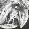
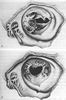
Mitral valve is a heart valve, which is composed
of two leaflets (anterior and posterior) demarcated by two commissures
along an annular ring, separating the left atrium from the left
ventricle, and serving as a door and guard for the entrance
of blood either from the left atrium or abnormally from the
left ventricle due to regurgitation. It opens and closes to
allow oxygenated blood to flow from the left atrium into the
left ventricle (see figures 104a,
104b,
105a,
105b,
106a,
106b).
Its leaflets are attached distally by fibrous strands called
chordae tendineae to papillary heart muscles in the cavity of
the left ventricle.
This valve's orifice or door may become too
narrow (stenosis, see figure 20a, 20b, 20c, 44a, 44b, 44c, 44d,
44e) due to rheumatic fever sequelae or congenital heart disease
including the rare deformity of the "parachute" mitral
valve (figure 44
g-1 and figure 44g-2)
or too wide to close properly (regurgitation, see figure 45,
69, 109, 118) due to various diseases, such as myocardial infarction
(heart attack, see definition of myocardial
infarction), infection (48b, 48h), degeneration, myxomatous
changes (see figure 115), congenital or genetic causes (see
definition congenital
heart disease in adults).
The mitral stenosis and/or regurgitation may
be treated with percutaneous balloon valvotomy (valvuloplasty)
or open heart surgery (see figure 109), or valve replacement
(see figure 49, 69, 107, 114).
EVALUATION AND MANAGEMENT OF CHRONIC
MITRAL REGURGITATION
There about 500,000 discharge diagnoses of mitral
valve disease annually in the United States. However, the estimates
of the prevalence of mitral regurgitation are confounded by
the presence of benign flow murmurs in many adults and by the
small amount of physiologic regurgitation detected on echocardiography
in 80% of adults. Only about 18,000 patients undergo mitral
valve surgery annually, suggesting that most patients with the
diagnosis of mitral regurgitation never need surgical intervention.
Thus, the challenge for the clinician is first to determine
which patients have the pathologic mitral regurgitation and
then to provide them with appropriate care.
THE CLINICAL PROBLEM
Causation
Normal mitral valve function depends on the
complex interactions of all the components of the valve apparatus
( Fig. 1 ). In surgical series, the most common causes of mitral
regurgitation are mitral-valve prolapse ( 20 to 70% of cases),
ischemia steamy and ( 13 to 30 %) rheumatic heart disease (3
to 40 percent), and endocarditis (10 t0 12%). Although mitral-
valve prolapse , is common in surgical series, most patients
with mitral- valve have only mild disease and never need surgery.
Mitral-valve prolapse and ischemic disease are also common in
patients with milder regurgitation , but the most common causes
are ventricular dilatation and systolic dysfunction. In the
elderly, mitral regurgitation may be due to annular calcification;
typically regurgitation in older people is mild to moderate
and intervention is rarely necessary. Accurate identification
of the mechanism of mitral regurgitation is essential because
the clinical outcome, the medical therapy prescribed and the
potential need of surgical intervention depends as much on the
cause as on the severity of the disease.
Pathophysiologic Process
Chronic left ventricular volume overload as
a result of mitral regurgitation lead compensatory dilatation
of the left ventricle. Although this response initially maintains
cardiac output, myocardial decompensation eventually results
in symptoms of heart failure and increased risk of sudden death.
In some patients, left ventricular contractility is an irreversibly
impaired in the absence of symptoms. In addition, backflow into
the left atrium results in the enlargement of the left atrium,atrial
fibrillation and elevated pulmonary pressures.
Diagnosis
Mitral regurgitation may be diagnosed on the
basis of the presence of a systolic murmur in asymptomatic adults
or incidentally when echocardiography is performed for other
indications. Some patients with primary disease of the valve
leaflets present with symptoms of heart failure, atrial fibrillation,
or endocarditis. The symptoms may be precipitated by a superimposed
hemodynamic stress, such as that induced by a pregnancy, anemia,or
an infection. In patients with secondary regurgitation, valve
dysfunction is most often identified during an evaluation of
the underlying disease process.
On physical examination, the murmur of mitral
regurgitation is classically apical holosystolic murmur that
radiates to the axilla. However, physical examination is not
always reliable in distinguishing mitral regurgitation from
other types of systolic murmurs and does not provide an accurate
measure of the severity of regurgitation. On electrocardiography
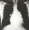
and chest radiography, evidence of enlargement of the left atrium,
left ventricle, or both is seen only late in the course of disease
and is not sensitive or specific for the diagnosis of mitral
regurgitation .
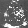
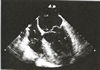
Echocardiography
Echocardiography allows accurate evaluation
of the presence or absence, severity and cause of mitral regurgitation.
Echocardiography is indicated in patients who have a systolic
murmur and any cardiac symptoms, a loud murmur (approximately
grade 3/6) alone, or other cardiac findings on physical examination.
In most cases, the cause of mitral regurgitation can be deduced
from the two dimensional images ( Fig. 202 B). Although Doppler
echocardiography provides several methods of quantifying the
severity of regurgitation, none have been shown to predict the
clinical outcome. Most centers grade regurgitation as mild,
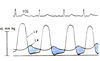
moderate or severe using a combination of color flow,continuous,
and pulse-wave Doppler imaging.
The most important aspect of the echocardiographic
examination is the quantitation of left ventricular systolic
performance. Although calculation of the ejection fraction is
an imperfect means of assessing contractility, from a practical
point of view the ejection fraction in conjunction with the
end systolic dimension provides a clinically useful measure
of ventricular performance. Transesophageal echocardiography
allows accurate assessment of the feasibility of valve repair
and should be performed before surgical intervention.
Outcome
Patients with mitral regurgitation may remain
asymptomatic for many years; the average interval from diagnosis
to the onset of symptoms is 16 years. There are few data on
the rate of hemodynamic progression of disease in patients with
mild to moderate regurgitation, since most series are restricted
to patients with severe regurgitation. In addition, the available
data are difficult to interpret, since the criteria for evaluating
the severity of regurgitation vary and are not always clearly
defined. Furthermore, even though the clinical outcome is strongly
dependent on the cause of the disease, patients with diverse
mechanisms of regurgitation are often included in the same study.
In patients with severe symptomatic mitral regurgitation,
the clinical outcome is poor: survival rates are as low as 33%
at eight years in the absence of surgical intervention.The average
mortality rate is approximately 5percent per year; most deaths
are related to heart failure, but there is a substantial incidence
of sudden death, suggesting that ventricular arrhythmias may
be an important feature of the disease process. Other complications
include atrial fibrillation,cerebral ischemic events, and endocarditis.
In patients with mitral-valve prolapse the
clinical outcome depends on the extent of the leaflet disease
and the severity of mitral regurgitation. The progression of
disease may be slow and insidious or maybe abrupt, as a result
of a chordial rupture leading to the flail leaflet in one study
of patients with initially asymptomatic severe mitral regurgitation
caused by mitral-valve prolapse, only 28% required surgery within
five years because of the onset of symptoms. In contrast, 90
percent of patients with flail mitral-valve leaflet died or
underwent surgery within 10 years, whether or not they initially
had symptoms.
Mitral regurgitation as a sequelae of rheumatic
fever is uncommon in the United States and is typically associated
with some degree of mitral stenosis. Ischemic mitral regurgitation
encompasses several mechanisms, including papillary muscle dysfunction,
regional ventricular dysfunction, and left ventricular dilatation.
The outcome is related to the severity of symptoms at presentation
and the extent of underlying coronary disease. In patients with
dilated cardiomyopathy, mitral regurgitation has diverse causes,
including annular dilatation, changes in the shape and size
of the left ventricle and systolic dysfunction.
STRATEGIES AND EVIDENCE
Most patients in whom chronic mitral regurgitation
is diagnosed have mild to moderate disease and are unlikely
to ever need surgical intervention. Management is directed toward
identifying the cause and severity of the regurgitation, treating
underlying disease processes, preventing complications, educating
the patient ,and evaluating risk factors for coronary disease.
In patients with primary mitral-valve disease periodic echocardiography
allows early detection of impaired left ventricular systolic
function on the basis of measurement of the end systolic dimension
and ejection fraction ( table 1 ). Other echocardiographicm
measures that are useful in clinical decision making include
assessment of the size of the left atrium and pulmonary systolic
pressure .
Medical Therapy
No known medical therapies directly affect
the disease process in the valve leaflets in patients with mitral-valve
prolapse or rheumatic valve disease. There has been sustained
interest in the concept of using vasodilator therapy to decrease
severity of mitral left ventricular dilatation. The rationale
for vasodilator care is that a reduction in the after load may
increase aortic flow and decrease mitral backflow. To some extent,
this rationale has been validated in small, short term studies
that have demonstrated a decrease in systemic vascular resistance
and regurgitant fraction and an increase in cardiac output with
vasodilator therapy, often with a decrease in ventricular volume
and end-diastolic pressure. However, these studies show that
vasodilators are most effective in improving symptoms in patients
with mitral regurgitation associated with ventricular and impaired
systolic function. There are no data that support the use of
vasodilator therapy in patients with asymptomatic mitral regurgitation
and normal ventricular function. Most important, the use of
medical therapy should not delay consideration of surgical intervention
in patients with symptoms or evidence of left ventricular systolic
dysfunction. Medical therapy is primarily directed toward the
treatment of complications of mitral regurgitation and the prevention
of endocarditis with antibiotic prophylaxis. If atrial fibrillation
occurs, standard approaches to rate control, cardioversion and
anticoagulation are indicated. In patients with mitral regurgitation
as result of ischemic disease prevention of ischemia with medical
therapy, percutaneous transluminal coronary intervention or
bypass grafting is appropriate. In patients with mitral regurgitation
due to dilated cardiomyopathy, medical therapy for heart failure
including afterload reduction,often results in improvement in
left ventricular shape, size and systolic function in association
with a reduction in the severity of regurgitation .
Mitral-Valve Surgery
The optimal surgical intervention for mitral
regurgitation is valve repair. As compared with valve replacement,
successful valve repair results in superior hemodynamics and
ventricular function, avoidance of a prosthetic valve and the
need for long term anticoagulation, and less distortion of ventricular
shape. The feasibility of valve repair is highest in patients
with mitral- valve prolapse, especially in those whose disease
is confined to the posterior leaflet. As surgical techniques
improve, an increasing number of patients are becoming candidates
for this procedure. When valve repair is not technically possible,
every effort is made to maintain the integrity of mitral chordal
apparatus. With chordal preservation, there is little change
in the ejection fraction after surgery, as compared with an
average decline of 10 ejection- fraction units in patients with
transected chords. The operative mortality is lower for mitral-valve
prolapse than for valve replacement (2 to 4% versus 5 to 10%)
In patients with mitral-valve prolapse, long term. clinical
outcome is excellent, with survival rates of 80 to 94% at 5
to 10 years with valve repair as compared with 40 to 60% with
valve replacement.
In patients with symptoms due to the mitral
regurgitation, surgical intervention is indicated, unless they
have severe left ventricular dysfunction. In asymptomatic patients
with severe mitral regurgitation, the outcome is improved if
surgery is performed before the onset of irreversible ventricular
dysfunction. No randomized trials have assessed the optimal
timing of intervention for asymptomatic severe mitral regurgitation,
and the ideal measure of ventricular contractility remains elusive.
However, a consensus has been reached that left ventricular
end-systolic dimensions and ejection fraction can be used to
identify the early systolic dysfunction. The evidence supporting
this approach is derived from studies in patients who were undergoing
valve surgery for severe mitral regurgitation that assessed
the value of preoperative variables as predictors of postoperative
ventricular performance. Indicators of early systolic dysfunction
are an end systolic dimension of 45mm or more or an ejection
fraction of 0.60 or less. Systolic dysfunction is most likely
when both valves are abnormal and sequential studies show a
progressive deterioration. Other factors that may affect the
timing of surgical intervention include the feasibility of valve
repair, the onset of atrial fibrillation, and the development
of pulmonary hypertension ( Fig. 3
). There are two noteworthy features of these criteria: the
degree of ventricular dilatation seen with isolated volume overload
due to mitral regurgitation is much less than that seen in aortic
regurgitation ,a condition characterized by combined pressure
and volume overload, and these criteria only apply to patients
with severe mitral regurgitation.
AREAS OF UNCERTAINTY
Assessment of the Severity of Mitral Regurgitation
The current definition of severe mitral regurgitation
is based on angiographic and echocardiographic descriptors of
the degree of backflow across the valve. An alternative physiological
definition would be mitral regurgitation severe enough to result
in dilatation of the left ventricle, left atrium or both. However,
the best left definition would be regurgitation leading to adverse
clinical outcomes. Unfortunately, prospective data based on
quantitative measures of severity are not unavailable.Thus,
it is not certain that some patients with moderate regurgitation
have severe disease that has not yet resulted in the ventricular
enlargement. The percentage of patients with mild regurgitation
will have a progressive increase in the severity of mitral regurgitation
is also unknown.
Medical Therapy for Primary Valve Disease
In patients with severe mitral regurgitation
due to primary valve disease, there are no persuasive data that
medical therapy decreases the rate of ventricular dilatation
or delays valve surgery. Some clinicians argue that medical
therapy may even be harmful if it is in creases the severity
of the regurgitation in patients with mitral-prolapse, prevents
normal adaptive responses of the left ventricle, or delays the
recognition of early symptoms or ventricular dysfunction.
Timing of Surgical Intervention
When severe mitral regurgitation and severely
reduced ventricular function are both present, it can be difficult
to determine whether ventricular dysfunction is the cause or
a consequence of chronic regurgitation. In either case, the
surgical outcome is poor when the ejection fraction is less
than 0.3, unless chordal continuity is preserved. In some patients,
a trial of medical therapy for heart failure and an evaluation
of the other causes of left ventricular dysfunction being clarify
the situation. Because the optimal approach to these patients
is controversial clinical decision making must be individualized
on the basis of the evaluation of the ventricular and valve
function, the likelihood of valve repair,the presence of other
underlying conditions and the patients'preferences.
Valve Repair in Patients with Secondary
Mitral Regurgitation
Some studies of patients with ischemic mitral
regurgitation suggest that revascularization alone decreases
the serverity of regurgitation,whereas other studies suggest
that concurrent valve repair or the placement of an annuloplasty
ring is necessary. Revascularization might be effective if regurgitation
is due to ischemia or if revascularization improves the shape
of the mitral valve. However,if there is irreversible myocardial
damage or if remodeling does not occur, then mitral regurgitation
may persist. In the absence of randomized clinical trials the
surgical decision is currently individualized on the basis of
mechanism of regurgitation in each patient.In patients with
dilated cardiomyopathy ,mitral regurgitation is due to change
in the shape of the valve apparatus , so that the severity of
regurgitation is often decreased by medical therapy that restores
ventricular size and shape .Some centers advocate mitral valve
sugery inthese patrients,but this approach is not widely accepted.
GUIDELINES
The American College of Cardiology and the
American Heart Association have developed detailed guidelines
for evaluation, follow-up, and optimal timing of surgical intervention
in patients with severe mitral regurgitation. Appropriate candidates
for mitral-valve surgery include patients with symptoms, except
those with severe ventricular dysfunction and in patients with
no symptoms who have mild to moderate ventricular dysfunction.
Surgery is indicated in asymptomatic patients with preserved
ventricular function if there is ahigh likelihood of valve repair
or if there is evidence of pulmonary hypertension or recent
atrial fibrillation.Guidelines also address the use of echocardiography,
the prevention of rheumatic fever and endocarditis, and indications
for anticoagulation.
CONCLUSIONS AND RECOMMENDATIONS
In the case of patients with a cardiac murmur,
the threshold for echocardiographic evaluation should be low.When
the valve is anatomically abnormal, the periodic clinical an
echocardiographic follow-up allows early identification of symptoms,
complications, and systolic dysfunction. In patients with secondary
mitral regurgitation, echocardiography serves as a first step
toward the evaluation and treatment of the underlying disease
process. Patient education is vital, both to ensure compliance
with follow-up and allow the patient to participate in the decision
making process. Surgical intervention in patients with severe
mitral regurgitation is indicated at the onset of symptoms or
in the presence of convincing evidence of left ventricular dysfunction.
Valve repair rather than valve replacement should be performed
whenever possible. We should remain cautious in recommending
valve surgery for asymptomatic patients who are considered to
have severe regurgitation but who have no evidence of consequences
of hemodynamic abnormalities. However, the excellent anatomical
and clinical outcomes of valve repair make surgical intervention
appropriate earlier in the course of disease in many patients
with severe mitral regurgitation as a means of preventing chronic
volume overload.
Otto,C.M.,Evaluation and Management of Chronic
Mitral Regurgitation, N Engl J Med,Vol.345,No 10,9/6/2001,PP.740-746.
| Mitral
Valve Percutaneous Balloon Valvotomy (PMV) |
This
is a procedure in which a cardiac catheter containing a balloon
expanding device is inserted percutaneously through the right
femoral vein and on into the right side of the heart. This device
has a cutting mechanism (i.e. needle) which allows the catheter
to be passed through the interatrial septum (see figures 35a,
104b)
and thus into the left atrium.
From there the balloon mechanism can be advanced through the
mitral valve and inflated accordingly to treat the mitral stenosis.
The
balloon catheter can also be advanced with the help of a guide
wire out the aortic valve into the ascending aorta. In order
to pass various larger dilating balloon catheters across the
atrial septum and the stenotic mitral valve, the hole in the
interatrial septum may need to be dilated as well. The balloons
are ultimately positioned to straddle the mitral valve and one
or two simultaneous inflations are performed. (see figure
69, 108).