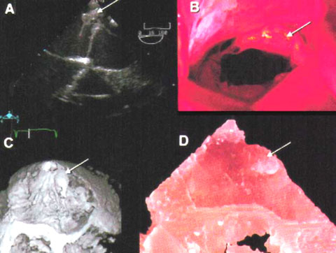
Figure 20c
This illustration show 1) the abnormality of the mitral valve called flail posterior leaflet obtained with a transesophageal echocardiography instrument (see white arrow). Also shown 2) is a mitral valve prolapse (see figure 45) (in which the leaflet protrude abnormal back into the left atrial chamber during systole when the mitral valves closes (arrow). There is a chordal rupture (which is a tearing of the tissue called chordae tendinae (figure104b) anchoring the mitral valve to muscle wall projections in the cavity of the left ventricle, as well as the flail posterior mitral leaflet (medial scallop).
Reference:Binder,T.M.,Moertl,D.,Mundigler,G.,Rehak,G.,Frankee,M.,Delle-Karth,G.,Mohl,W.,Baumgartner,H.,Mauer,G., JACC,vol.35,Jan. pages230-237,modified