


The most common infiltrative cardiomyopathy is amyloidosis, which by electron microscopy consists of rigid, nonbranching fibrils lying in between the myocardial muscle cells (see figures 74c, 74d, 77b). There are two kinds of amyloidosis:
1) primary
in which there is no coexisting disease, except in some cases
of multiple myeloma (a disease of the plasma cell, which proliferates
out of control to crowd out the normal bone marrow cells resulting
in anemia and other abnormalities).
2) secondary amyloidosis may be associated with chronic diseases
like tuberculosis, or rheumatoid arthritis. The heart becomes
stiff as a result of the infiltration of the amyloid into the
heart muscular tissue leading to restrictive heart mechanisms
of contraction.
|
 Figure 74a |
 Figure 74b |
 Figure 74c |
 Figure 74d |
 Figure 77a |
 Figure 77b |
 Figure 77c |
 Figure 77d |
 Figure 77e |


The echocardiogram shows thickening of both the right and left ventricular muscle (to see the entire photographs of figures above click on: 73a, 73b, 74a, 74b, 74c, 74d, 77a, 77b, 77c, 77d, 77e).
The prognosis is poor and most treatments are ineffective.
In addition to an EKG, chest x-ray, and Doppler echocardiogram, cardiac catheterization is necessary to document the diagnosis with special attention being focused on the venous pressure, which is characteristically elevated considerably above normal, with an initial dissent and then a plateau.
While the left systolic ventricular pressure is normal,the left ventricular diastolic pressure shows the same abnormalities as the right ventricular blood pressure (see figures 74a, 77d, 77e).
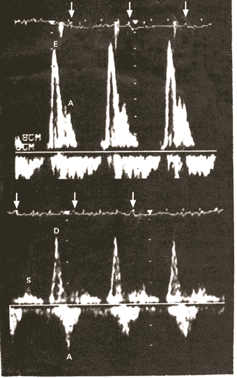
Figure 74a Pulsed Doppler Flow Patterns of Transmitral Inflow (Upper Panel) and Pulmonary Venous Inflow (Lower Panel), with Characteristic Features of Restrictive Cardiomyopathy (in this case due to amyloid deposition).
The upper panel shows a prominent early diastolic filling wave (E) with rapid deceleration and a smaller atrial filling wave (A). The lower panel shows blunted pulmonary venous inflow in systole (S), with a prominent diastolic filling wave (D), which decelerates rapidly, followed by a reversal of flow into the pulmonary veins in late diastole during atrial contraction (A). The arrows in both panels show the R wave of the electrocardiogram (EKG).
This illustration demonstrates the use of the Doppler echocardiogram to study blood flows in and out of various chambers of the heart to make diagnoses.
DiSalvo, T.G., King, M.E., Smith, R.N., Case Records of the Massachusetts General Hospital, A 66-Year-Old Woman with Diabetes, Coronary Disease, Orthostatic Hypotension, and the Nephrotic Syndrome, NEJM, Vol. 342, Jan 27-00, p 264-274. (modified)