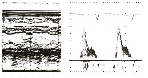
Figure 77a
M-mode echocardiogram showing increased thickness of LV wall, consistent with LV hypertrophy, and on the right a Doppler tracing of mitral inflow. Note that velocity is maximum at middiastole. The atrial contribution to mitral blood flow velocity is considerably reduced.
Shabetai, R., MD, Restrictive Cardiomyopathy, Hurst's The Heart, 8th edition, p 1643. (modified)