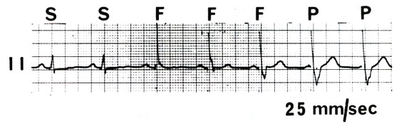
Figure 89
Electrocardiogram lead II demonstrating normal ventricular inhibited (VVI) pacing. The first two complexes are sensed (S) and there is pulse generator output inhibition. The last two complexes show ventricular pacing (P). Complexes 3 to 5 are fusion beats (F), although complex 3 may more correctly be called a pseudofusion beat as the stimulus artifact is late and does not contribute to the QRS. This can be clearly seen by observing the T-wave changes from the sinus complexes through the fusion beats to the paced beats.
Mond, H.G., MD, Permanent Cardiac Pacemakers: Techniques of Implantation, Testing, and Surveillance, Hurst's The Heart, 8th ed., p 815-841