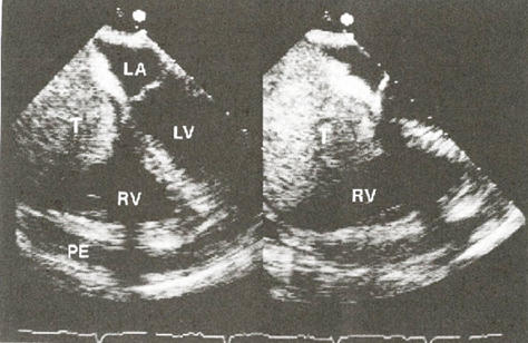
Figure 72b
Transesophageal echocardiogram in a 55-year-old woman who presented with adenocarcinoma of the lung and obstructed superior vena cava syndrome. A large tumor (T) is seen in the right ventricle (RV) in systole (left panel) and diastole (right panel). Subsequent images revealed that it originated from an obstructed superior vena cava (which ordinarily receives venous blood from the arms, head, and neck). The echo-free space anterior to the right ventricle represented pericardial effusion (PE). LA, left atrium. LV, left ventricle.
Hall, R.J., MD, Cooley, D.A., MD, MCAllister, Jr., H.A., MD, Frazier, O.H., MD, Neoplastic Heart Disease, Hurst's The Heart, 8th edition, p 2020.