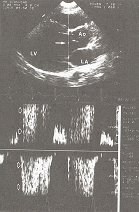
Figure 48i
Aortic regurgitation, pulsed wave. Doppler
recording.
Top: Parasternal long-axis image, showing orientation of the Doppler
cursor and location of the smple volume (arrow) in the center of the
outflow tract, on the left ventricular side of the aortic leaflets.
LV, left ventricle; Ao, aorta; LA, left atrium.
Bottom: Spectral Doppler recording. Diastolic regurgitant flow is disturbed,
and thus multidirectional, causing a broad spectrum of Doppler shifts
(open arrows) that last through diastole.