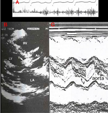
Figure 48a
Echocardiogram
from a patient with an aortic valve vegetation (infected mass of fibrin,
blood cells, platelets, etc., as a result of a blood stream infection).
A. Two-dimensional parasternal long-axis view shows a very dense, nodular
structure (arrow v) that represents a large vegetation. This diastolic
frame shows the vegetation projecting into the left ventricle (LV).
B. M-mode study shows a shaggy, dense structure in diastole (arrow v)
that represents the aortic valve vegetation. Ao, aorta; LA, left atrium;
RV, right ventricle.
J.M. Felner M.D., R.P. Martin M.D., The Echocardiogram, The Hurst's The Heart, 8th ed., p 407. (modified)