Figure 46a
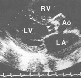
Two-dimensional echocardiogram from a patient with severe calcific aortic stenosis. Parasternal long-axis view in systole shows dense masses of calcium (arrowheads) representing the anterior (right coronary) and posterior (noncoronary) cusps virtually obscuring the leaflets and limiting their excursion. Ao, aorta; RV, right ventricle; LV, left ventricle; LA, left atrium.
J.M. Felner, M.D., R.P. Martin,
M.D., The Echocardiogram, The Hurst's The Heart, 8th ed., p 406.