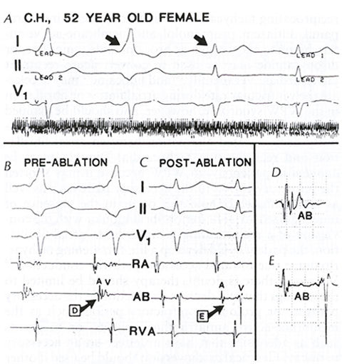
Figure 3b
Radiofrequency
ablation (the technique involves heart catheterization with a catheter
containing a wire capable of delivering radiofrequency energy to selected
areas in the cardiac conductive system) in WPW syndrome. The patient
had frequent recurrent supraventricular tachycardias due to WPW syndrome.
A. Standard leads I, II, and V1 demonstrate disappearance of
the delta wave from one impulse to the next, 5s after beginning the
application of radiofrequency energy (compare successive QRS complexes
indicated by arrows).
B. Prior to ablation, the interval between atrial (A) and ventricular
(V) activation at the site of the ablation catheter is below 50 ms,
and the sharp spike between A and V likely represents activity in the
bypass tract.
C. Immediately after ablation, the A-V interval at the site of
the ablation catheter (AB) is lengthened to 150 ms, and the accessory
pathway spike has disappeared.
D and E. Enlargements of D and E from panels B and C, respectively.
(RA= right atrium, AB= ablation catheter, RVA= right ventricular apex.)
Myerburg, R.J., MD, Kessler, K.M., MD, Castellanos, A., MD, Recognition, Clinical Assessment, and Management of Arrhytmias and Conduction Disturbances, Hurst's The Heart, 8th edition, p 705-758.