Figure
39c
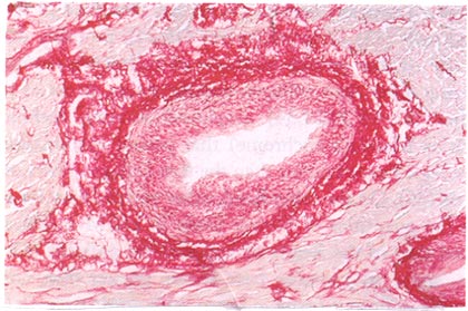
High-power view of a transversely cut abnormal intramural coronary artery in a ventricular septal tissue section from a 28-year-old patient with HCM. Picrosirius red stain shows dense perivascular (adventitial) collagen, as well as increased amounts of collagen in the thickened media. (Magnification x100)
Shirani, J., MD, Pick, R., MD, Roberts, W.C., MD, Maron, B.J., MD, Morphology and Significance of the Left Ventricular Collagen Network in Young Patients With Hypertrophic Cardiomyopathy and Sudden Cardiac Death, Journal of American College of Cardiology, Vol 35, No 1, 2000, p 41.