Figure
38e
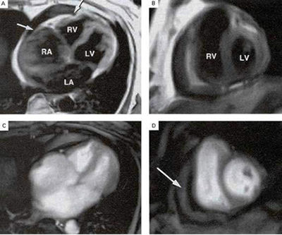
Constrictive Pericarditis
With
marked neck vein distention, swelling of the legs, and a knocking noise
over the heart. Chest films showed extensive calcifications on the anterior
and inferior surfaces of the heart involving both layers of pericardium.
Magnetic resonance images (MRI), in panel A and panel B, show diffuse
thickening of the entire pericardium, calcifications (arrows
in Panel A), right atrial (RA) enlargement, a moderate amount
of fluid between the two pericardial layers, and increased signal in
the cavity, indicative of stasis of blood.
Panels C and D confirm the presence of pericardial fluid (arrow
in Panel D) and showed atrial enlargement as well as an abnormal
"shivering" motion of the interventricular septum. Pericardiectomy showed
partially calcified tissue. RV denotes right ventricle, LV left ventricle,
and LA left atrium.
Benedetti, E.D., Didier, D., Constrictive Pericarditis, The New England Journal of Medicine, July 13, 2000, p 107.