figure
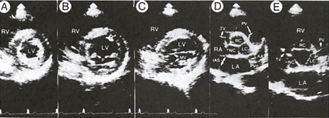
34a
Two-dimensional
parasternal short-axis views from a normal subject
A. The level of the cardiac apex shows a small, thick-walled left ventricle
(LV) and only a very small portion of the right ventricle (RV).
B. Left ventricle at the level of the papillary muscles (P). The right
ventricle is larger and the left ventricular cavity is uniformly round
and larger than at the apical level.
C. Left ventricle, at the level of the partially opened mitral valve
(MV), is larger than at the papillary muscle level.
D. Diastolic frame at the base fo the heart shows the closed aortic
valve, with the right (RC), left (LC), and noncoronary (NC) cusps resembling
the letter Y. The tricuspid (TV) and pulmonic (PV) valves are identified.
E. Systolic frame at the same level as D shows the open aortic valve.
IAS, interatrial septum; LA, left atrium; RA, right atrium; LAA, left
atrial appendage; PVn, pulmonary vein.
J.M. Felner, R.P. Martin, The Echocardiogram, The Hurst's The Heart, 8th ed., p 388.