Figure
33
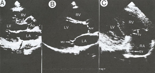
Two-dimensional
parasternal long-axis views from a normal subject.
A.
Long-axis plane of the left ventricle (LV) in diastole. The aorta (Ao)
and the left atrium (LA) are to the right of the sector image, and the
apex of the left of the sector image. The outflow tract portion of the
right ventricle (RV) appears anteriorly, at the top or narrowest part
of the fan-shaped image; the posterior walls of the left ventricle and
left atrium appear near the bottom or widest portion of the image. The
coronary sinus (CS) and descending aorta (Dao) are the most posterior
structures.
B.
Enlargement of view A in systole excludes the most posterior structures
and show that the leaflets of the aortic valve (AoV) open to the periphery
and the leaflets of the mitral valve (MV) are closed. The left ventricle
is two to three times the size of the right ventricle.
C.
Right ventricular inflow tract view shows the right atrium (RA), right
ventricle, tricuspid valve (TV), and a small portion of the left ventricle.
J.M. Felner, R.P. Martin, The Echocardiogram, The Hurst's The Heart, 8th ed., p 387.