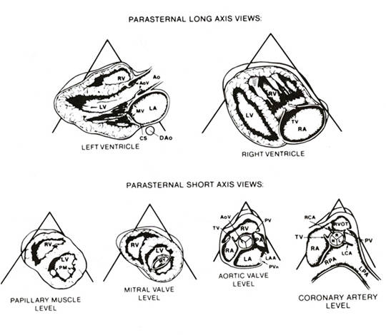
Figure
32d
Anatomic drawings of the parasternal imaging planes oriented as they would be seen on the video screen. The two long-axis views illustrate the plane of the left ventricle (LV) and surrounding structures and the plane of the right ventricular inflow tract. The four short-axis views illustrate the left ventricle at the papillary muscle (PM) and mitral valve (MV) levels and the levels of the great vessels and coronary arteries. RV, right ventricle; AoV, aortic valve; Ao, aorta; LA, left atrium; CS, coronary sinus; Dao, descending aorta; TV, tricuspid valve; PA, right atrium; PV, pulmonic valve; PA, pulmonary artery; LAA, left atrial appendage; PVn, pulmonary vein; RCA, right coronary artery; LCA, left coronary artery; R, right coronary cusp; L, left coronary cusp; N, noncoronary cusp; RVOT, right ventricular outflow tract; RPA, right pulmonary artery; LPA, left pulmonary artery.
DJ Sahn, R. Anderson: Two-Dimensional Anatomy of the Heart: An Atlas for Echocardiographers. John Wiley & Sons, N.Y., 1982.