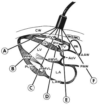
Figure
32c
Cross
section of the heart.
Lines A through F show how the ultrasound beam
is swept between the apex and the base of the heart.
With the transducer at A, the ultrasound beam traverses the skin-transducer
interface, chest wall(CW), anterior right ventricular wall (ARVW), right
ventricle (RV), interventricular septum(ISV), left ventricle(LV) at
the apex, and posterior left ventricular wall (PLVW). As the transducer
is tilted progressively cephalad, the following structures are imaged:
at B, the left ventricle at the posterior papillary muscle (PPM);
at C, anterior (AMVL) and posterior (PMVL) mitral valve leaflets;
at D, anterior mitral leaflet at the junction of the posterior left
ventricular wall and the left atrial wall (LAW) ;
at E, tricuspid valve (TV), anterior (AAW) and posterior (PAW) aortic
walls, aortic valve (AoV), and left atrium (LA);
at F, pulmonary artery (PA).
JM Felner,RC Schlant:Echocardiography:A Teaching Atlas.NY,Grune and Stratton,1976.