Figure
32a
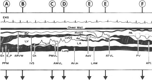
Representation
of an M-mode echocardiographic recording as the ultrasonic beam is swept
in an arc from the apex to the base of the heart. The arrows labeled
A through F refers to various positions of the ultrasound transducer
on the chest, over the heart. An ECG is shown for timing. The ultrasound
beam traverses the following structures:
at A, the homogeneous, stationary echo of the chest wall (CW), anterior
right ventricular wall (ARVW), right ventricle (RV), muscular portion
of the interventricular septum (IVS), left ventricle (LV) at the level
of the posterior papillary muscle (PPM), free left ventricular wall
myocardium, consisting of endocardium (En) and epicardial (E) and pericardial
(P) interface and lung;
at B, left ventricle at the level of the chordae tendineae (ch);
at C, the right and left ventricles at the level of the anterior (AMVL)
and posterior (PMVL) mitral valve leaflets;
at D, the left ventricular outflow tract (LVOT) anteriror to the mitral
valve;
at E, the right ventricular outflow tract (RVOT), ascending aorta (Ao),
aortic valve (AoV), left atrium (LA), and left atrial wall (LAW);
at E, the right venticle, anterior tricuspid valve leaflet (ATVL), right
atrium (RA), and left atrium;
at F, the pulmonary artery (PA), pulmonic valve (PV), atriopulmonic
sulcus (APS), and left atrium.
JM Felner, RC Schlant: Echocardiography: A Teaching Atlas. New York, Grune & Stratton, 1976.