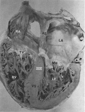
Figure 24c
Four-chamber view of heart showing morphologic differences between the four chambers. The right atrium (RA) is more trabeculated than the left (LA), and the right ventricle (RV) is more heavily and coarsely trabeculated compared to the left ventricle (LV). AS= atrial septum; MV= mitral valve; TV= tricuspic valve; VS= ventricular septum.
From BF Valler et al: Tomographic
views of normal and abnormal hearts: The anatomic basis for various
cardiac imaging techniques. Clin Cardiol 13:804(pt I), 877(pt II), 1990