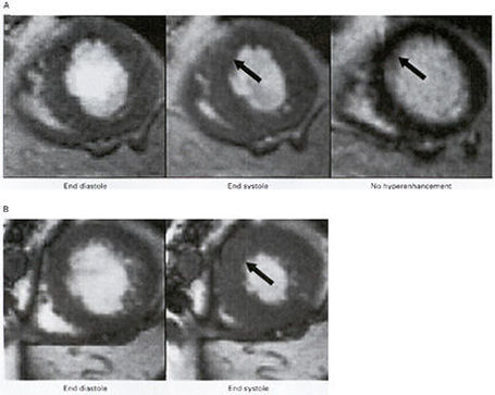
Figure124
Representative Cine Images and Contrast-Enhanced Images Obtained by MRI in One Patient with Reversible Ventricular Dysfunction (Panels A and B) and One with Irreversible Ventricular Dysfunction (Panels C and D in Figure125). The patient with reversible dysfunction had severe hypokinesia (reduced contraction of the involved myocardium) of the anteroseptal wall (arrows), and this area was not hyperenhanced before revascularization. The contractility improved after revascularization.The patient with irreversible dysfunction had akinesia (no motion of the involved myocardium) of the anterolateral wall (arrow), and this area was hyperenhanced before revascularization. The contractility of the wall did not improve after revascularization (Figure125)
Reference:R.Kim and others.The New England Journal of Medicine, Vol.343, Nov.16, 2000,No.20,Pp.1445-1453.Modified