
Figure 120a
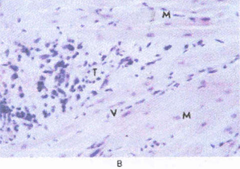
Figure120b
Panel A shows a trans esophageal echocardiogram (horizontal view) at the level of the left ventricular outflow tract. A tumor is attached to the left atrial (LA) side of the anterior mitral leaflet (thin arrow), and there is a smaller mass at the ring insertion of the posterior leaflet (thick arrow).
Examination of the resected mass (Panel B) showed a mesenchymal, poorly differentiated tumor (R), consisting of polymorphic, predominantly spindle-shaped cells spreading within the myocardium (M) along preformed vessels (V), corresponding to an angiosarcoma (hematoxylin and eosin, x200). LV denotes left ventricle, and Ao aorta. (see definition of Echocardiogram)
Dennig, K., MD, Lehmann, G., MD, Richter, T., MD, The Angiocarcoma in the Left Atrium, The New England Journal of Medicine, Vol. 342, Jan 13-00, p 443-444.