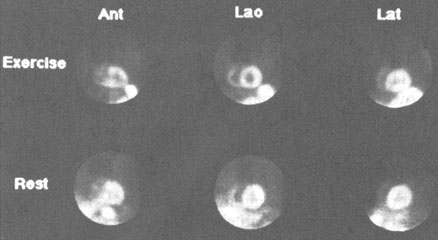
Figure 109c
Three view (ungated) planar 99mTc-sestamibi scans performed following injection at peak exercise. Note the clear visualization of the right ventricular free wall. There are septal and apical defects on the LAO and ANT views, respectively, which demonstrate fill-in on the "Rest" injected images (bottom row).
Johnson, L.L., MD, Pohost, G.M., MD, Nuclear Cardiology, Hurst's The Heart, 8th edition, p 2281-2295.