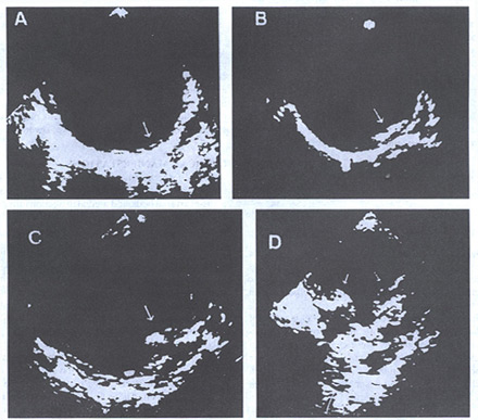
Figure 105d
Panels from transesophageal echocardiographic examinations of the descending thoracic aorta in four patients. Varying degrees of atherosclerotic plaquing are illustrated. A minimal degree is shown in A (upper left) and a more severe but still moderate degree in B (upper right). C and D demonstrate protruding plaques, the configuration with the most serious threat of embolization.
(From Lindsay J.Jr et al. Diseases of the aorta. In: Schlant RC, Alexander AW, Lipton MJ, eds. Diagnostic Atlas of the Heart. New York: McGraw-Hill, 1996:319. Reproduced with permission from the publisher and authors.)