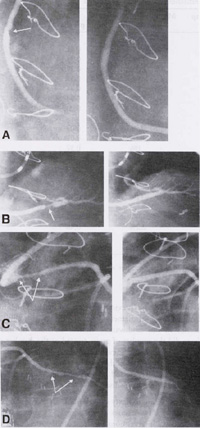
Figure 95b
A. Before (arrow, left panel) and after (right panel) stenting of the proximal SVG to the right coronary artery (RCA).
B. Before (arrow, left panel) and after (right panel) stenting of the distal SVG to the RCA.
C. Before (arrows, left panel) and after (right panel) stenting of diffuse proximal disease in the SVG to the left anterior descending coronary artery (LAD).
D. Before (arrows, left panel) and after (right panel) stenting of distal disease in the SVG to the LAD, with excellent angiographic results at both treatment sites.
All four SVG lesions were in the same patient and treated at the same intervention.
Bhargava, Balram, MD, et al., Multiple Saphenous Vein Graft Stenting, Journal of American College of Cardiology, Vol 35, No 2, Feb 2000, p 389-397.