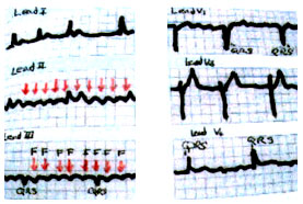
Figure 5a
Atrial Flutter.
Note the sawtooth pattern in Leads II and III, discrete F atrial waves in V1 and poorly registered atrial actively in I and V6.
Myerburg, R.J., M.D., Castellanos, A., M.D., Kessler, K.M., M.D., Recognition, Clinical Assessment and Management of Arrhythmias and Conduction Disturbances, Hurst's The Heart, 8th ed, p 722.