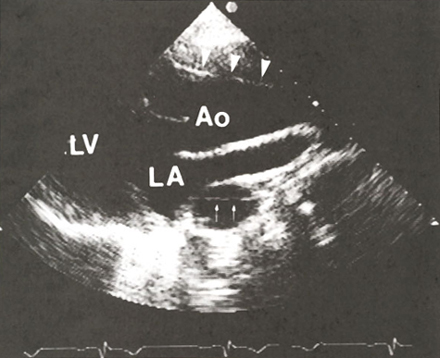
Figure 50
Two-dimensional
parasternal echocardiogram from a patient with a Type I (proximal) dissection
of the aorta.
The ascending aorta (Ao) is dilated and contains a linear density (arrowheads)
that represents the intimal (referring to the inner wall of the aorta)
flap, which separates the true and false lumen (opening).
The descending aorta is visible behind the left atrium (LA) and also
contains the intimal flap (small arrows). LV, left ventricle.
J.M. Felner M.D., R.P. Martin M.D., The Echocardiogram, The Hurst's The Heart, 8th ed., p 409. (modified)