Figure 4.vsd
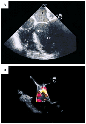
Transesophageal Four-Chamber Views
in a Patient with a Ventricular Septal Rupture Complicating an Acute
Posterior Myocardial Infarction.
In Panel A, the ventricular septal rupture (arrow) is basal and at the
junction between the basal inferior septum and the inferoposterior wall.
In Panel B, a color Doppler image shows the shunt across the ventricular
septal rupture. LA denotes left atrium, LV left ventricle, and RV right
ventricle.