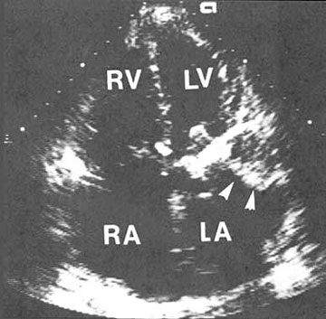
Figure 49
Two-dimensional apical four-chamber view from a patient with a prosthetic mechanical mitral valve. The left atrium (LA) is dilated and contains a dense echogenic structure (arrowheads) attached to the lateral wall in the region of the left atrial appendage that t represents a large left atrial thrombus (blood clot). A portion of the mitral valve prosthesis is seen projecting into the left ventricle (LV) and angling toward the intraventricular septum. LV, left ventricle; RA, right atrium; RV, right ventricle.
J.M. Felner M.D., R.P. Martin M.D., The Echocardiogram, The Hurst's The Heart, 8th ed., p 408. (modified)