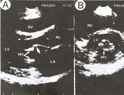
Figure 44a
Two-dimensional echocardiogram from a patient with moderate mitral stenosis. A. Parasternal long-axis view in diastole demonstrates doming (arrow) of the anterior leaflet, commonly seen in rheumatic mitral stenosis in the absence of marked valvular calcification. The leaflet tips are only moderately calcified, and the left atrium (LA) is moderately dilated. The extent of leaflet tip separation of the mitral valve (MV) is beneficial for identifying the true orifice in the parasternal short-axis plane and is also helpful as a rough guide to judge whether the parasternal short-axis view through the tips of the mitral leaflets demonstrates the technique for determining mitral valve area. Planimetry of the valve area (dotted line) is performed at the onset of diastole when the orifice appears the largest; the valve orifice measures 1.2 cm2. LV, left ventricle; RV, right ventricle; Ao, aorta.
J.M. Felner, M.D., R.P. Martin, M.D., The Echocardiogram, The Hurst's The Heart, 8th ed., p 404.