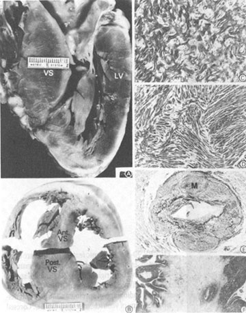
Figure 40b
Morphologic compounds of the underlying
disease process in HCM.
A. Gross heart specimen sectioned in a cross-sectional plane similar
to that of the echocardiographic (parasternal) long axis. The pattern
of left ventricular hypertrophy is asymmetrical, with wall thickening
confined primarily to the anterior ventricular septum (VS), which bulges
into the left ventricular outflow tract.
B. Heart specimen illustrating a different pattern of hypertrophy in
which marked left ventricular wall thickening is localized to the posterior
portion of the ventricular septum (Post. VS), while the anterior septum
(Ant. VS) is only mildly thickened.
C, D. Histology characteristic of the left ventricle in HCM. In C, septal
myocardium shows markedly disordered architecture with adjacent hypertrophied
cardiac muscle cells arranged at perpendicular and oblique angles to
each other. In D, bundles of hypertrophied cells show a disorganized,
interwoven arrangement.
E. Intramural coronary artery with apparently narrowed lumen and thickened
wall due primarily to medial (M) hypertrophy. F. Extensive scarring
of ventricular septum which is transmural in distribution. LV= left
ventricular free wall.
From BJ Maron et al: N Engl J Med 316:780,