Figure 40a
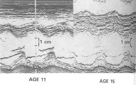
Development and progression of left
ventricular hypertrophy in children with HCM.
Development of marked hypertrophy of the anterior basal ventricular
septum (VS). M-mode echocardiograms shown here were obtained at the
same cross-sectional level in a girl with a family history of HCM. At
age 11, ventricular septal thickness was at upper limit of normal (10
mm); at age 15, septal thickness had increased markedly (to 33 mm),
and appearance of the echocardiogram is typical of HCM. The patient
remained asymptomatic throughout this period of time but died suddenly
and unexpectedly at age 17.
PW= posterior left ventricular free wall. BJ Maron et al: NEJM, 315:610, 1986.