Figure 3.vsd
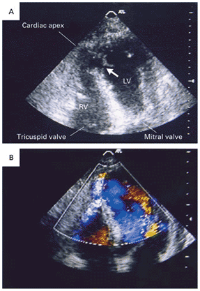
Transthoracic Two-Dimensional Apical
Four-Chamber Views in a Patient with Ventricular Septal Rupture Complicating
an Anterior Acute Myocardial Infarction.
In Panel A, the ventricular septal rupture is apical and simple (arrow).
RV denotes right ventricle, and LV left ventricle. The sites of both
the right and the left ventricular perforation are at the same level.
In Panel B, color Doppler ultrasonography shows turbulent flow (bright
blue mosaic) from the left ventricle to the right ventricle through
the interventricular septal defect.