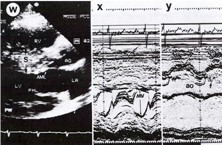
Figure 39i
Echocardiogram
from a patient with hypertrophic cardiomyopathy.
W. Two-dimensional parasternal long-axis
view in diastole demonstrates asymmetric septal hypertrophy. The thickness
of the ventricular septal wall (S) is 30 mm and that of the posterior
wall (PW) is 20 mm. Characteristic brightening of the septal echoes
is evident. The left ventricular outflow tract between the anterior
mitral leaflet (AML) and the septum is virtually obliterated. The left
atrium (IA) is dilated.
X. M-mode study at the level of the
mitral valve shows systolic anterior motion (arrow) of the mitral valve
and the mitral valve (MV) abuts against the intraventricular septum
in diastole.
Y. M-mode study at the level of the
aorta shows premature closure (arrow) of the anterior aortic leaflet.
PML, posterior mitral leaflet; Ao, aorta; RV, right ventricle.