 |
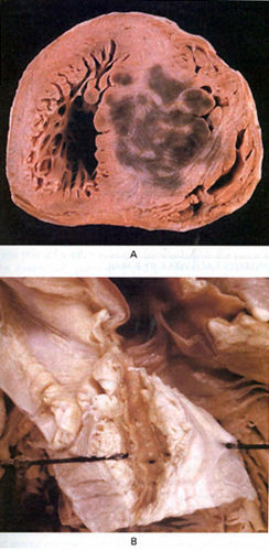
The Heart of a Boy with Hypertrophic Cardiomyopathy. |
|
Basso, C., MD, Thiene, G., MD, Gori, F., MD, Myocardial Infarction in a Patient with Hypertrophic Cardiomyopathy, NEJM, Vol 342, No 8, Feb 24, 2000, p 593. (modified) Figure 39f |
|
|
The Heart of a Boy with Hypertrophic Cardiomyopathy. |
|
Basso, C., MD, Thiene, G., MD, Gori, F., MD, Myocardial Infarction in a Patient with Hypertrophic Cardiomyopathy, NEJM, Vol 342, No 8, Feb 24, 2000, p 593. (modified) Figure 39f |
|