Figure
39d
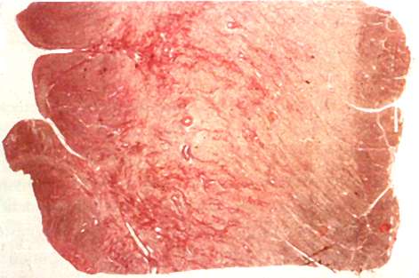
Low-power view of ventricular septal tissue section from a 20-year-old patient with HCM stained with picrosirius red showing widely distributed interstitial collagen. Volume fraction of interstitial collagen in this patient was 18%. Abnormal intramural coronary arteries with thickened walls are also visible. (Magnification x6)
Shirani, J., MD, Pick, R., MD, Roberts, W.C., MD, Maron, B.J., MD, Morphology and Significance of the Left Ventricular Collagen Network in Young Patients With Hypertrophic Cardiomyopathy and Sudden Cardiac Death, Journal of American College of Cardiology, Vol 35, No 1, 2000, p 41.