Figure
37
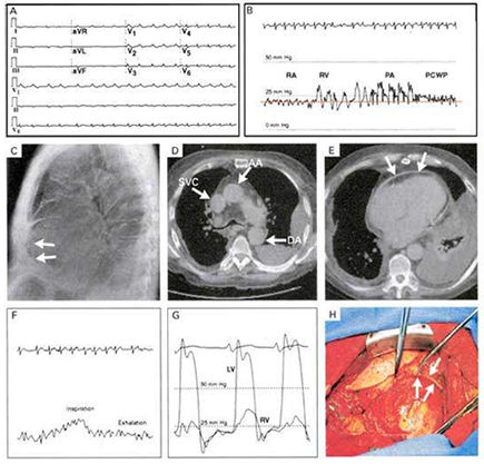
A
case of Constrictive Pericarditis with swollen legs, neck vein distension,
enlarged liver, and EKG showing low voltage and ectopic atrial tachycardia
(Panel A). Heart catherterization showed diastolic equalization of the
right atrial (RA) pressure, right ventricular (RV) pressure, pulmonary-artery
(PA) pressure, and pulmonary -capillary wedge pressure (PCWP), indicated
by the red line in Panel B.
Chest film showed a thickened pericardium (arrows in Panel C). Computed
tomograhpic scan (CT) showed a dilated superior vena cava (SVC), and
a thickened pericardium (arrows in Panel E). Heart catherterization
also showed dialstolic equalization of the left and right ventricular
pressures.
Marked pericardial thickening (P) (arrows in Panel H) was also shown
during pericardial stripping.
Atwood, J.E., Osterberg, L., Constructive Pericarditis, The New England Journal of Medicine, July 13, 2000, p106.