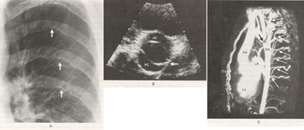
Figure 23b
Aortic Coarctation and Bicuspid
Aortic Valve
This is a case of a 30-year-old man being investigated for a bicuspid
aortic valve, chest x-ray showed marked rib notching (arrows in Panel
A), due to marked collateral channels from the axillary and internal
thoracic arteries. An echocardiogram confirmed a bicuspid aortic valve
without stenosis (Panel B), and continuous-wave Doppler scanning of
the descending aorta showed marked diastolic runoff consistent with
the presence of severe coarctation. An MRI of the chest showed severe
focal coarctation (black arrow in Panel C), measuring 1 mm, just beyond
the left subclavian artery (which takes off from the aortic arch and
goes to the left arm). Numerous large collaterals were present (white
arrows). AC denotes anterior cusp, PC posterior cusp, AA ascending aorta,
DA descending aorta, ITA internal thoracic arteries, LV left ventricle,
and LA left atrium.
Burce, C.J., MB, Breen, J.F., MD, Aortic Coarctation and Bicuspid Aortic Valve, The New England Journal of Medicine, Vol 342, Jan 27-00, p 249 (modified