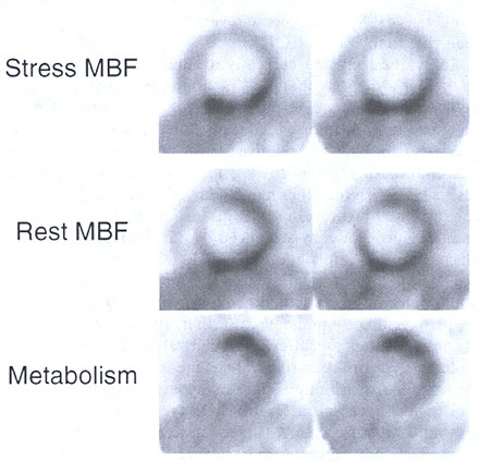
Figure 216-14
Two contiguous short-axis images through the mid-left ventricle in a patient with ischemic cardiomyopathy. The MBF images at rest (middle panel) reveal in the interventricular septum reduced but relatively welt preserved flow in the anterior and anterolateral wall associated with regionally increased ["F]deoxyglucose uptake in the same portion of the left ventricle as seen in the lower panel (metabolism). The stress induced flow defect in the anterior and anterolateral wall (as seen in the upper panel) implicates stunning as the cause of the enhanced ["F]deoxyglucose uptake.