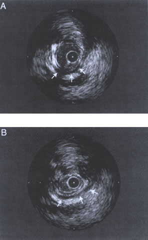
Figure 212-10
Intravascular ultrasound imaging of the myocardial bridging in diastole (A) and in systole (B). A typical half-moon-shaped echolucent area surrounds the bridge during the entire cardiac cycle (arrows). Note the catheter artifact in diastole (A) at seven o'clock.