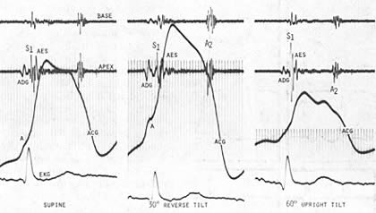
Figure 199d
Base and apexphonocardiograms are recorded simultaneously with the aid the apexcardiogram(ADG) and ECG in an 52 year old man with significant systemic hypertension. In all three postures an atrial diastolic gallop(ADG) precedes the mitral component of S1. In turn M1 is followed by a prominent second component that is most likely an aortic root ejection sound in this clinical setting. in the supine position S1 is of moderate intensity. during a 30° reverse tilt with the increased venous return, There is a significant increase in the amplitude of the ADG as well as prominence of the "a" wave of ACG. A DG is further separated from the mitral component of S1, which is slightly softer. In contrasts , with 60° of upright tilt and a decrease in venous return, there is a significant decrease in the intensity of the ADG and loss of the "a" wave on the AVG.note also the migration all of the ADG toward S1 which has increased significantly in intensity. With these postural changes there's no but significant change in the intensity of the aortic ejection sound(AES), which is a well recorded at the apex an at the base. These simple maneuvers at the bedside can be particularly helpful in distinguishing an (ADG-S1 sequence versus a split persist a split S1.