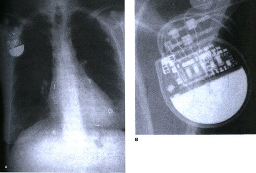
Figure 16d
A.
A chest x-ray of a patient with dual-chamber unipolar pacemaker. The leads
are placed inthe right atrial appendage and in the right ventricular apex,
respectively7. Note only one electrode at the distal tip of each lead.
B. Close
inspection of the pacemaker generator shows that the ventricular lead
has pulled out of the header with very minimal contact between the ventricular
electrodes and the metal contacts in the pacemaker header. This caused
intermittent failure to sense and pace in this patient.