Figure140
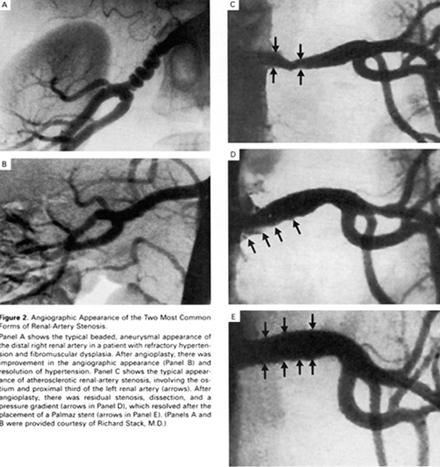
Angiographic Appearance of the Two Most Common forms of Renal-Artery Stenosis.
Panel A shows the typical beaded, aneurysmal appearance of the distal right renal artery in a patient with refractory hypertension and fibromuscular dysplasia.Afterr angioplasty, there was improvement in the angiographic appearance (Panel B) and resolution of hypertension. Panel C shows the typical appearance of atherosclerotic renal-artery stenosis ,involving the ostium and proximal third of the left renal artery(arrows). After angioplasty, there was residual stenosis ,dissection,and a pressure gradient(arrows in Panel D), which resolved after the placement of a Palmaz stent (arrows in Panel E).Panel A and B were provided courtesy of Richard Stack,MD)
Reference:Safian.R.and others.Renal Artery Stenosis,N.Engl.J.Med.Vol.344,No.6,Feb.8,2001.pp431-442