Figure
119
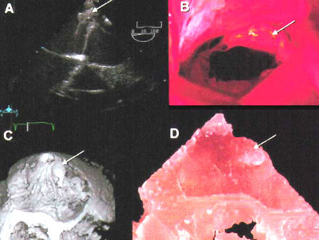
Flail posterior leaflet of the mitral valve: Flail leaflet (arrow) as seen during transesophageal echocardiography (A) intraoperative inspection of the valve (B) confirmed MV proplapse, chordal rupture and a flail posterior leaflet (medial scallop). The flail segment of the posterior leaflet can be appreciated in the 3D reconstruction (at systole) (C) and is also visible in the stereolithographic model (D).
Binder, T.M., Moertl, D., Mundigler, G., Rehak, G., Franke, M., Delle-Karth, G., Mohl, W., Baumgartner, H., Maurer, G., Stereolithographic Biomodeling to Create Tangible Hard Copies of Cardiac Structures from Echocardiographic Data: In Vitro and In Vivo Validation, Journal of the American College of Cardiology, Vol 35, Jan 2000, No 1, A-38.