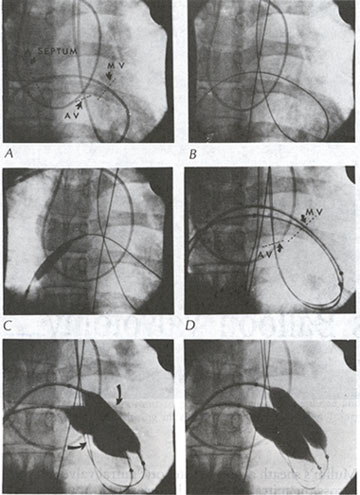
Figure 108
Sequence
of percutaneous mitral valvotomy.
A. Floating balloon catheter in position across the atrial septum through
the mitral and aortic valves. The tip is in the ascending aorta.
B. The 0.038-in transfer guide wire has been passed through the floating
balloon catheter. The floating balloon catheter has been removed.
C. An 8-mm dilating balloon catheter enlarging the atrial septal puncture
site.
D. Two 20-mm dilating balloon catheters advanced into position across
the stenotic mitral valve over two separate 0.038-in transfer guide wires.
E. Partially inflated dilating balloon catheters across the mitral valve.
Note the "waist" produced by the stenotic vlave (arrows).
F. Fully inflated dilating balloon catheters in position across the mitral
valve. (A. septum= atrial septum; AV=aortic valve; MV= mitral valve.)
Gaasch, W.H., M.D., O'Rourke, R.A., M.D., Cohn, L.H., M.D., Rackley, C.E., M.D., Mitral Valve Disease, Hurst's The Heart, 8th edition, 1994, p 1568.