These arrhythmia are usually persistent and are called nonparoxysmal.
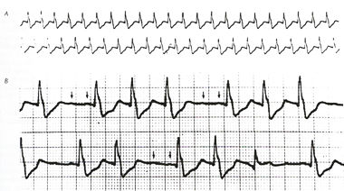
A.
A 20-s continuous recording demonstrates a regular tachycardia
at a ventricular rate of approximately 140 per minute.
B. During carotid sinus massage, ventricular
conduction becomes irregular and diminutive P waves
at twice the basic ventricular rate are evident Impaired
AV conduction with little or no effect on atrial activity
is characteristic of the response of an ectopic atrial
tachycardia to carotid sinus massage.
|
They may be due to reentry or automaticity mechanisms, or could
be due to triggered activity.
Often
a toxic or metabolic cause can be found responsible for the
tachycardia.
But focal atrial disease may play a part.
Digitalis in toxic amounts can cause such an arrhythmia with
a rate of 160 to over 200 per minute associated with a 2:1 conduction
or variable block (see illustration below).
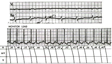
A.
Atrial tachycardia with 2:1 AV block due to digitalis
intoxication (note diminutive P waves, barely visible
even in V1)
B. Multifocal atrial tachycardia.
Note the constantly changing form of ectopic P waves.
Myerburg,
R.J., MD, Kessler, K.M., MD, Castellanos, A., MD, Recognition,
Clinical Assessment, and Management of Arrhytmias and
Conduction Disturbances, Hurst's The Heart, 8th edition,
p 705-758.
|
Decompensated chronic lung disease, acute alcohol abuse, metabolic
factors, electrolyte imbalances, and reduced oxygen blood levels
(hypoxemia) can cause similar arrhythmias.
Also, acute myocardial infarction (heart attack) can be a cause,
as well as trauma from prior open heart surgery.
The main treatment involves the correction of above causative
factors (when identifiable).
Since
atrial disease is often multicentric, surgical excision is not
often used.
Antiarrhythmic drugs may help if there is no identifiable cause.


